3/6/23
Breast cancer accounts for the greatest incidence of new cases of cancer and cancer-related deaths among women worldwide. Millions of women are diagnosed with breast cancer every year throughout the world, but although this is a disease that disproportionately affects female-identifying individuals, it is important to recognize that breast cancer has no gender. Wherever someone falls on the gender spectrum, there is a finite breast cancer risk that they and their care providers should understand. There are countless factors that cause someone to be at risk for breast cancer, including the organs they are born with, the organs they have at the time of cancer progression, lifestyle, and personal health history. Further, breast cancer’s metastatic nature means that it is transferable to distant organs, such as the bone, liver, lung, and brain. It is widely known that prevention and early diagnosis of the disease can lead to a good prognosis with a high survival rate. Thus, screening for breast cancer has been widely promoted, especially in high income countries (HICs). The three most common screening modalities are mammography (MMR), clinical breast examination (CBE), and breast self-examination (BSE).
In HICs, most recommendations for breast cancer screening have become synonymous with MMR, as it is the current standard for the screening and detection of breast cancer. MMR is a specific type of breast imaging that uses low-dose x-rays to see inside the breasts. During the exam, a radiology technician will position a patient’s breast on a special platform, where it will be compressed with a clear plastic paddle. This is necessary to even out the breast thickness so all the tissue can be visualized and small abnormalities are not hidden by overlaying tissue. Once the x-ray images are obtained, a radiologist interprets the exams by viewing and analyzing the images.
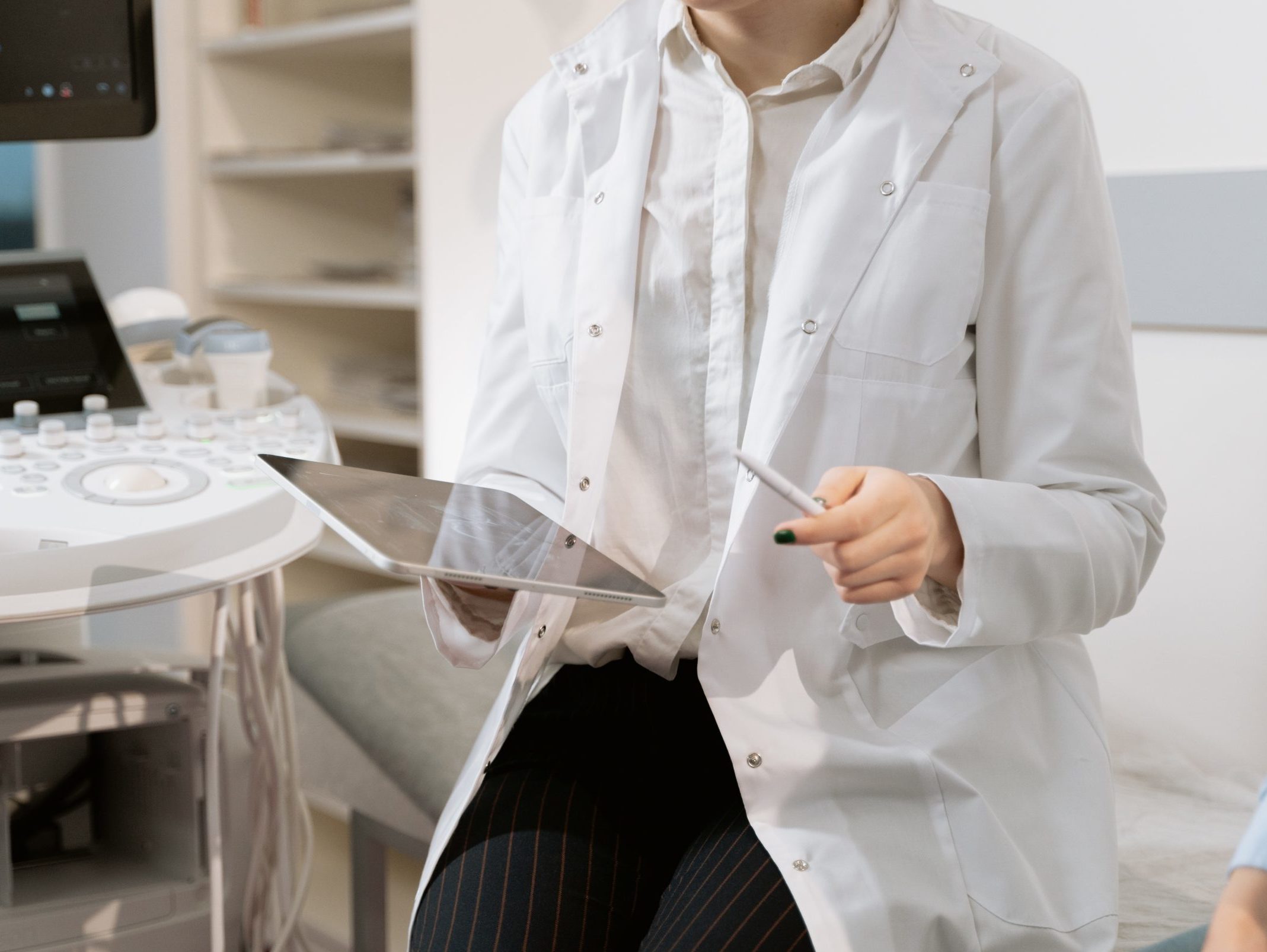
With the ever-growing wave of artificial intelligence (AI), several clinics and hospitals around the globe have started to experiment with using this technology to help radiologists detect breast cancer. In Hungary, which has one of the most robust breast cancer screening programs in the world, at least five hospitals or clinics have used AI programs since 2021. Successes in Hungary have prompted physicians in several other countries to experiment as well. In a 2022 study, it was found that an AI program that looked at 250,000 breast cancer scans was just as, if not more, effective than a human radiologist at detecting breast tissue abnormalities. The program was also able to view more images more quickly overall. The researchers concluded that incorporating this technology into the radiology field had the potential to heavily reduce the workload of physicians by providing an automated system that could provide a second opinion extremely efficiently.
While there are growing concerns that the use of this kind of technology could replace the role of a physician, it is important to understand that further rigorous clinical trials are needed before the systems can be more widely adopted and fully integrated into the healthcare system. The creators of AI cancer detection software have faced a great amount of scrutiny from doctors and health institutions, so there is still quite a ways to go before a patient can walk into a clinic and ask, “Is there AI involved in my breast exam?”
Nevertheless, quite a few physicians have been quite welcoming to this technology. With the long hours that many have to work, they recognize that there is room for human error or missed readings, which could be alleviated with the help of AI. However, one thing is for certain: an AI-plus-doctor should replace a doctor alone, but AI should never replace a doctor.

